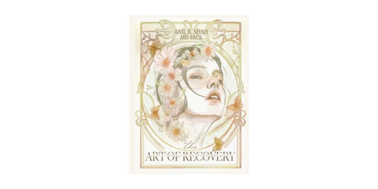
Anil R. Shah, MD, Eugene J. Kim, MD, Corey S. Maas, MD
Pathogenesis of Facial Aging
Facial aging generally consists of intrinsic and extrinsic processes. Intrinsically, the most extensive changes occur in the dermis, especially in its upper third. The total amount of ground substance, predominantly made of glycosaminoglycans and proteoglycans, diminishes. In addition, elastic fibers, which maintain the wavy pattern of collagen bundles, become thin and fragmented with time, beginning at the age of 30. Finally, the ratio of type I to type III collagen diminishes over time, along with a general reduction in the total amount of collagen.
Extrinsically, one of the main factors in facial aging is from actinic damage. In a process called elastosis, collagen is degraded and elastin is increased. The dermis becomes thickened and poorly organized. Fine-skin wrinkling from sun-damaged epidermis also occurs. Along with fat atrophy in the subcutaneous tissue, there is a general redistribution of tissue due to gravitational effects. The sagging of tissue leads to the loss of the cervicomental angle and mandibular definition.
Anatomy
Fascial Layers of the Face
The fascia of the face is complex; complete understanding is required in order to perform surgery of the face safely. The superficial musculoaponeurotic system (SMAS) is a distinct layer just superficial to the parotid fascia.(Figure 71–1). Generally, sensory nerves lie superficial and facial nerve fibers lie deep to the SMAS. Superiorly, the SMAS is contiguous with the temporoparietal fascia. Anteriorly, insertion occurs at the lateral border of the zygomaticus muscle and to the dermis of the upper lip. Thus, the posterior traction of the SMAS will actually deepen the nasolabial fold. The SMAS is also in continuity with the orbicularis oculi muscle in the lower eyelid. Inferiorly, there is continuity with the platysma over the body of the mandible and extension in the neck.
The superficial temporal fascia, also known as the temporparietal fascia (TPF), is located immediately below the subcutaneous tissues of the temporal area and is continuous with the SMAS of the lower face and the galea of the forehead. Beneath the TPF lies the deep temporal fascia. It splits at the level of the supraorbital ridge to become the intermediate temporal fascia and the deep temporal fascia. The intermediate fat pad lies beneath the intermediate temporal fascia, while the deep temporal fat pad lies beneath the deep temporal fascia.
Muscular Anatomy of the Face
The platysma is a broad, thin muscle innervated by the cervical branch of the facial nerve and is continuous with the SMAS of the face. Platysmal bands can form, along with pseudoherniation of fat, resulting in the loss of the cervicomental angle (“turkey gobbler”). The depressor muscles of the forehead are the corrugator supercilli, orbicularis oculi, procerus, and depressor supercilli. The lone elevator of the brow is the frontalis muscle. The corrugator produces vertical rhytids, while the procerus and frontalis muscles produce transverse and horizontal rhytids respectively. The frontalis muscle does not have a bony origin or insertion point (originates from the galea and inserts onto the orbicularis oculi). The corrugator muscle has an oblique head, which depresses the brow, and a transverse head, which pulls the brow medially with contraction. Excessive resection of the transverse head of the corrugator in certain patients can lead to unnatural widening of the brows.
Motor Nerves of the Face
The facial nerve (cranial nerve VII) gives mimetic function through innervation of the facial musculature. Divisions include the frontal, zygomatic, buccal, mandibular, and cervical branches (Figure 71–2). Crossover innervation occurs up to 70% in the zygomatic and buccal branches and only up to 15% in the frontal and mandibular branches. The frontal branch runs within the temporoparietal fascia and lies just superficial to the superficial layer of the deep temporal fascia. It generally crosses the zygoma with multiple branches approximately 4 cm lateral to the lateral canthus, halfway between the root of the helix and the lateral canthus. The Pitanguy method predicts the course of the nerve by drawing a line .5 cm inferior to the tragus to 1.5 cm above the lateral brow. The course of the facial nerve can also be estimated by drawing a line from the tragus to the lateral canthus and bisecting it with a line from the earlobe superiorly. The medial zygomaticotemporal vein (aka sentinel vein) is an important landmark of the location of the temporal branch of the facial nerve in endoscopic browlifts. The zygomatic and buccal branches are deep to the SMAS but become more superficial as they course anteriorly. The marginal branch has a varying course but generally extends up to 1 cm below the inferior edge of the mandible. All muscles of the face are innervated on their deep surface with the notable exceptions of the mentalis, levator anguli oris, and buccinator. In general the surgeon should be deep to the facial nerve in the upper face and superficial to the facial nerve in the lower face.
Sensory Nerves of the Face
The greater auricular nerve provides sensation to the inferior lobule and the upper neck and is a branch of the cervical plexus. The skin and SMAS lie just superficial to the nerve. The nerve has an intimate association with the sternocleidomastoid muscle and has been shown to cross the muscle at its midpoint, approximately 6 cm below the external auditory canal. The supratrochlear nerve emerges deep to the eyebrow and penetrates the corrugator muscle. The supraorbital nerve has a superficial branch (medially, passes over frontalis muscle) and a deep branch (laterally, passes between the galea and pericranium).
Ligaments of the face
The zygomatic ligament (MacGregor’s patch) and the mandibular ligament are true (connection between skin and bone) facial ligaments. Release of these ligaments is necessary to allow for adequate redraping of the face in a tension free manner.
Patient Selection
The appropriate selection of patients has critical importance in assuring a desirable outcome. Patients need to understand the goals and limitations of rhytidectomy. They should be motivated and should be willing to participate in other associated changes that support longevity, such as improved diet, smoking cessation, and sun protection.
Dedo developed a classification of the aging neck which accounts for skin, muscle, and fat redundancy and mandible and hyoid position. Patients with the hyoid located superiorly and posteriorly are ideal candidates for maximal cervicomental angle improvement.
Patient History & Physical Examination
Preparation for surgery should include a complete history and physical examination, including bleeding history and hypertension. Patients should cease smoking and the use of nonsteroidal anti-inflammatory medications approximately 2 weeks prior to and after surgery.
Pre-existing brow asymmetries should be brought to the patient’s attention prior to any surgical manipulation of the brow. Ideally, the female brow should have a peak above the orbital rim at the lateral limbus. Hairline and forehead shape should be evaluated to determine candidacy for brow procedures. Significant brow asymmetries respond best to direct brow lifts, especially if there is preexisting rhytid on the forehead. High hairlines and sharply sloped foreheads are best treated with a pretrichial incision, to prevent posterior hairline displacement.
Rhytidectomy
Surgical Techniques
Incisions
The goals of incision placement are to minimize hair loss, hairline adjustments, visible scars, and changes in normal anatomic structures. Pretragal incisions avoid the need to redrape hair-bearing skin onto the tragus with the disadvantage of a more visible scar. Post-tragal incisions are hidden behind the tragus, but require attention to recreate the normal contour of the tragus. If the temporal area requires lifting, the incision is curved anterosuperiorly into the hair-bearing scalp. If alopecia is a concern, the incision is brought more anteriorly in a pretrichial or “trichophytic” fashion to preserve the patient’s hairline (Figure 71–3).
The posterior incision is preferably brought into the hair if minimal skin redraping is necessary. If there is a large amount of anticipated redraping of the skin posterior, the incision can be placed along the hairline to circumvent standing cutaneous deformities.
Subcutaneous Rhytidectomy
The subcutaneous flap is elevated with facelift scissors just superficial to the SMAS using a combination of traction and countertraction (Figure 71–4). Elevation of the flap into the neck extends to the middle third of the neck. The risk of facial nerve injury increases once the anterior border of the parotid gland is reached. Superiorly, dissection is carried in a plane superficial to the temporalis fascia, which allows the protection of the frontal facial nerve branch. The risks of hematoma, skin flap ischemia, and facial nerve injury increase in longer skin flaps. In patients who may be at risk for skin flap ischemia (ie, smokers) a shorter skin flap may be desirable. Subcutaneous rhytidectomy generally provides greater tension on incisions resulting in wider facial scars and limited facial rejuvenation.
SMAS Rhytidectomy
Suture plication or the infolding of the SMAS eliminates the need to incise the SMAS, while imbrication involves undermining and cutting the SMAS prior to suspension. Studies have shown no difference in aesthetic outcome in patients with imbrication versus plication. The SMAS is incised in the preauricular and neck regions and elevated using blunt dissection. The limits of the flap are just below the zygomatic arch superiorly, the anterior border of the parotid gland anteriorly, and the angle of the mandible inferiorly (Figure 71–5). Techniques that address the SMAS-platysma complex have been shown to have more favorable long-term results as compared to skin flap elevation alone. SMAS techniques provide rejuvenation of the neck and jowls with no improvement in nasolabial folds.
Deep-Plane & Composite Rhytidectomy
Deep-plane rhytidectomy attempts to address aging changes in the melolabial fold and malar area. Composite rhytidectomy essentially extends the deep plane by including the orbicularis oculi muscle in the dissection. Essentially, a composite flap is created, which is preplatysmal in the neck, suborbicularis oculi in the malar regions, and sub-SMAS anterior to the cheek fat pad. (Figure 71–7). The deep plane and composite rhytidectomy release facial ligaments and therefore provide maximal redraping of the skin and provide the least amount of tension on the external scar. Studies have failed to show a significant difference in effacement of nasolabial folds between deep plane techniques and SMAS techniques.
Closure & Drains
The direction of suspension for the skin flap is posterior and superior. Making the majority of the closure tension free is critical. The points of maximal tension are in the temporal and occipital regions and key sutures are placed to suspend the skin flap. Following suspension, excess skin is trimmed so that the skin edges are reapproximated in a tension-free manner.
The pretragal area is carefully defatted and a subcutaneous anchoring suture is placed to recreate the normal concave contour of this area. Closed-suction drainage is placed through a separate stab incision in the hair-bearing skin; however, no studies show that drains actually help prevent hematomas. Drains do allow for the removal of residual fluid and help to keep the skin flap adherent to the underlying tissue. The use of tissue sealants that use fibrin, thrombin, or platelet-rich gel (or any combination of these substances) may obviate the need for drains.
Postoperative Care
The wounds are cleaned and antibiotic ointment is placed on the incisions. Bulky dressings are placed immediately following the procedure and help in keeping pressure on the skin flap. The head of the bed is kept elevated and patients are placed on limited bed rest. Blood pressure control with the patient’s normal medication, along with the control of pain and nausea, are extremely important in the immediate postoperative period.
Complications
Nerve Injury
The most commonly injured nerve in rhytidectomy is the greater auricular nerve, with a 6% rate of injury. Facial nerve injury is relatively rare and has been reported to be 2.6% with subcutaneous techniques and up to 4.3% in sub-SMAS techniques. The most commonly injured motor branch of the facial nerve is the temporal branch.
Hematoma
Hematoma is the most common complication of rhytidectomy and has an occurrence rate of 1–8%. Men are twice as prone to developing hematoma secondary to the increased vascularity in the hair-bearing skin of the face. Unilateral pain or pain unresponsive to medication needs an immediate evaluation for the possibility of a hematoma. Hematoma is a worrisome complication because of the risk of epidermolysis and skin slough. Symptoms and signs of hematoma include an abrupt increase in pain, swelling, and ecchymosis, which are especially concerning if they are unilateral. Rapidly expanding hematomas necessitate a return to the operating room and the control of bleeding points. When hematomas are delayed or small, they can be managed with needle aspiration and pressure dressings. The incidence of hematoma can be minimized with careful intraoperative hemostasis, blood pressure control, and the cessation of NSAIDs.
Additional Complications
Other complications of rhytidectomy include scarring, a “pixie ear” deformity, an elevated temporal hairline, and skin slough. These can be prevented by careful incision placement and minimizing tension on the suture line. Infection is a relatively rare complication of rhytidectomy and is treated with antibiotic therapy.
Baker D. Rhytidectomy with lateral SMASectomy. Facial Plast Surg. 2000;16(3):209. [PMID: 11802569] (Review of the SMAS excision technique.)
Baker DC, Conley J. Avoiding facial nerve injuries in rhytidectomy. Anatomical variations and pitfalls. Plast Reconstr Surg. 1979;64(6):781. [PMID: 515227] (A classic review of facial nerve anatomy in the context of rhytidectomy.)
Brennan HG, Toft KM, Dunham BP, Goode RL, Koch RJ. Prevention and correction of temporal hair loss in rhytidectomy. Plast Reconstr Surg. 1999;104(7):2219. [PMID: 11149791] (Review of decision making in approaching the temporal hair tuft.)
Grover R, Jones BM, Waterhouse N. The prevention of haematoma following rhytidectomy: a review of 1078 consecutive facelifts. Br J Plast Surg. 2001;54(6):481. [PMID: 11513508] (Retrospective analysis of the risk factors for hematoma.)
Hamra ST. The deep-plane rhytidectomy. Plast Reconstr Surg. 1990;86(1):53. [PMID: 2359803] (Classic article first describing the deep-plane rhytidectomy.)
Hamra ST. Composite rhytidectomy. Plast Reconstr Surg. 1992; 90(1):1. [PMID: 1615067] (Classic article first describing the composite rhytidectomy.)
Kamer FM. One hundred consecutive deep plane face-lifts. Arch Otolaryngol Head Neck Surg. 1996;122(1):17. [PMID: 8554742] (Prospective study of the deep-plane rhytidectomy.)
Kamer FM, Frankel AS. SMAS rhytidectomy versus deep plane rhytidectomy: an objective comparison. Plast Reconstr Surg. 1998;102(3):878. [PMID: 9727459] (Retrospective review that favors the deep-plane rhytidectomy.)
Larrabee WF Jr, Henderson JL. Face lift: the anatomic basis for a safe, long-lasting procedure. Facial Plast Surg. 2000;16(3): 239. [PMID: 11802572] (Anatomic review and its implications in successful rhytidectomy.)
McCollough EG, Perkins SW, Langsdon PR. SASMAS suspension rhytidectomy: rationale and long-term experience. Arch Otolaryngol Head Neck Surg. 1989;115(2):228. [PMID: 2643976] (A substantial retrospective review of the SMAS technique by an expert on rhytidectomy.)
Mitz V, Peyronie M. The superficial musculo-aponeurotic system (SMAS) in the parotid and cheek area. Plast Reconstr Surg. 1976;58(1):80. [PMID: 935283] (The classic article first describing the SMAS anatomically.)
Perkins SW, Williams JD, Macdonald K, Robinson EB. Prevention of seromas and hematomas after face-lift surgery with the use of postoperative vacuum drains. Arch Otolaryngol Head Neck Surg. 1997;123(7):743. [PMID: 9236595] (Retrospective review showing the effectiveness of drains in decreasing seroma and hematoma formation.)
Sherris DA, Larrabee WF Jr. Anatomic considerations in rhytidectomy. Facial Plast Surg. 1996;12(3):215. [PMID: 9243990] (Review article of relevant anatomy.)
Browlift
Endoscopic Browlift
The endoscopic browlift is the currently the most commonly performed procedure for rejuvenation of the brow. The ideal candidate is a female patient with a normal or low hairline, thin skin, and a flat forehead. The endoscopic browlift can be performed in either a subgaleal or subperiosteal plane. Releasing the periosteum of the arcus marginalis and the orbital ligament adequately mobilizes the brow and helps achieve a long term result. The method of fixation of the brow varies with polylactic resorbable screws or bone tunnels. Some surgeons have found achieved long term brow elevation without the need for fixation. The endoscopic forehead lift has the advantage of minimal incision length and less risk of paraesthesias.
Coronal Browlift/Tricophytic Browlift/ Pretrichial Browlift
In a coronal browlift, the surgeon makes an incision within the hair-bearing scalp. The procedure will lead to elevation of the hairline. The plane of dissection is in the subgaleal plane allowing access to the frontalis, corrugator, and procerus muscles. Coronal brow lifting is contraindicated in patient’s with high hairlines or male pattern baldness. In addition, paraesthesias are common after this technique due to resection of the deep branch of the supraorbital nerve.
The tricophytic browlift makes an incision within the fine hair of the anterior hairline. A reverse bevel allows for hair growth through the incision. A pretrichial variant places the scar in front of the hairline. Both approaches are effective in patients with high hairlines.
Mid-forehead Brow Lift/Direct Browlift
A mid-forehead browlift is made with an incision in a horizontal furrow in the forehead. A direct brow lift is made by directly excising skin above a patient’s brow. Typical candidates for both procedures include bald male patients with prominent forehead wrinkles and patients with a highly asymmetric brow (facial paralysis patients). Forehead wrinkles and corruagtor hyperactivity are not addressed with this technique.
Midface lift
Midface lifts are techniques designed to specifically address the malar fat pad. A variety of approaches can be used including temporal (via an endoscope) with or without buccal release, and transconjuctival. A suture or polylactic acid device engages the malar fat pad and lifts it in a posterior superior direction, effacing the nasolabial fold. The suture is then fixed to deep temporal fascia. If a transconjuctival approach is utilized, fixation can occur by drilling a polylactic acid screw within the anterior face of the maxilla. When the nasolabial fold is lifted superiorly, excess skin will bunch along the lower lash line. Typically, the excess skin is resected in a conservative fashion. Patients who are candidates are generally younger patients who have isolated aging in the midface without prominent jowls or signs of aging present in the neck.
Complications
The most common complications of forehead lifting are residual brow asymmetry, hematoma, paraesthesias, and temporary paresis of the temporal branch of the facial nerve. Permanent paralysis of the temporal branch is rare. Poor scarring can result with excessive tension on surgical wounds or cauterization of hair follicles. Alopecia can be minimized with limited cautery and atraumatic tissue handling.
Midface lift patients may experience prolonged swelling due to disruption of lymphatics of the midface. Eye shape may change to appear, cat-like, but usually resolves with time. Preservation of a cuff of periosteum along the lateral orbit minimizes this complication. Excessive skin excision may lead to ectropion, especially if the midface returns to its preoperative level.
Schmidt BL, Pogrel MA, Hakim-Faal Z: The course of the temporal branch of the facial nerve in the periorbital region. J Oral Maxillofac Surg 2001 Feb; 59(2): 178-84
Cook TA, Brownrigg PJ, Wang TD, Quatela VC: The versatile mid-forehead browlift. Arch Otolaryngol Head Neck Surg 1989 Feb; 115(2): 163-8
Sykes JM: Surgical rejuvenation of the brow and forehead. Facial Plast Surg 1999; 15(3): 183-91
Trinei FA, Januszkiewicz J, Nahai F: The sentinel vein: an important reference point for surgery in the temporal region. Plast Reconstr Surg 1998 Jan; 101(1): 27-32
Knize DM: A study of the supraorbital nerve. Plast Reconstr Surg 1995 Sep; 96(3): 564-9
Figure 71–1. The SMAS is a distinct layer between the parotid gland, skin, and subcutaneous fat.
Figure 71–2. Facial nerve anatomy.
Figure 71–3. Possible incisions in rhytidectomy.
Figure 71–4. Elevation of the skin flap in the subcutaneous plane.
Figure 71–5. Incision of the SMAS.
Figure 71–6. Suspension of the SMAS superiorly and posteriorly.
Figure 71–7. Composite rhytidectomy with dissection of the orbicularis oculi and zygomaticus muscles.