
Posted by Dr. Anil R. Shah
Choosing to have rhinoplasty is a personal decision, and one of the most important parts of the process is making sure you and your surgeon have a clear understanding of your goals.
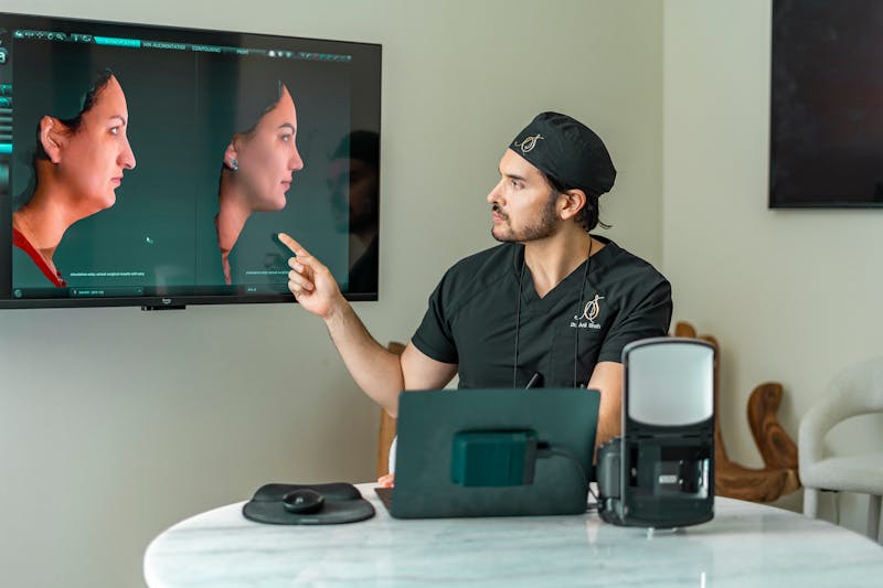
One technology that has transformed the rhinoplasty consultation is 3D facial imaging.
Unlike traditional photographs, 3D imaging allows both the patient and surgeon to look at the nose and face from multiple angles and explore potential changes in a more interactive way.
What Is 3D Facial Imaging?
Many modern 3D imaging systems use a technique called stereo photogrammetry.
The system captures photographs from multiple directions and uses fixed facial landmarks to create a three-dimensional representation of the patient's face.
The result is essentially a digital 3D model that can be viewed and manipulated from different angles.
Some systems can achieve sub-millimeter accuracy under controlled conditions, although the exact accuracy depends on the technology and how the imaging is performed.
Why Is 3D Better Than Looking at 2D Photos?
Traditional photographs are still an important part of a rhinoplasty consultation, but they have limitations.
A photograph provides a single view of the face. You can look at the nose from the front, side, or another angle, but each image is ultimately a separate two-dimensional representation.
With a 3D model, the surgeon can evaluate the nose more holistically.
Think of it like the difference between looking at a photograph of a sculpture and actually being able to rotate the sculpture in your hands.
The surgeon can manipulate the digital model and evaluate how a proposed change affects the nose from multiple perspectives.
Understanding Volumetric Changes
Some 3D imaging systems include tools that allow the surgeon to calculate and visualize volumetric differences between the existing nose and a proposed digital modification.
These changes can be displayed on the model as a visual overlay, allowing the surgeon to see where volume would theoretically be added or removed.
This can provide another layer of information when discussing a patient's goals.
Importantly, these measurements are part of the planning process—not a guarantee of what will happen during surgery.
How Does the Technology Help With Surgical Planning?
A patient's desired result is only one part of the equation.
A surgeon also has to consider the patient's existing anatomy, skin thickness, cartilage, bone structure, previous surgeries, and other factors that can influence the final result.
For example, the thickness and elasticity of the skin can affect how much an underlying structural change will actually be visible.
By combining the patient's aesthetic goals with an understanding of their anatomy, the surgeon can determine what changes may be realistic.
3D Imaging Can Also Improve Communication
Perhaps one of the biggest benefits of 3D imaging is communication.
Patients sometimes have difficulty describing exactly what they like or dislike about their nose.
Being able to manipulate a 3D model can give patients a visual way to communicate their preferences.
A patient might say they want a straighter profile, a more refined tip, or a subtle change in projection. The 3D model can help the surgeon understand what the patient means and discuss what may or may not be achievable.
This doesn't mean the digital simulation becomes a surgical blueprint.
Instead, it's a communication and planning tool.
Can Your Surgical Result Look Exactly Like the 3D Simulation?
This is one of the most important things to understand.
No digital simulation can guarantee an exact surgical outcome.
Surgery takes place on living tissue, and healing is influenced by factors that cannot be perfectly predicted. Swelling, scar formation, skin thickness, tissue elasticity, and individual healing can all affect the final result.
The simulation should therefore be viewed as a representation of a potential direction, rather than a promise.
Setting Realistic Expectations
The goal of 3D imaging isn't to promise perfection.
It's to help the surgeon understand what you want, evaluate what may be realistically achievable, and develop a surgical plan that is appropriate for your anatomy.
The technology can make the consultation more interactive and precise, but it doesn't replace the surgeon's experience or clinical judgment.
The Bottom Line
3D imaging has become a valuable tool in modern rhinoplasty consultations.
By creating an interactive model of the patient's face, it allows surgeons and patients to discuss potential changes from multiple angles and can provide additional information about the proposed changes.
Most importantly, 3D imaging can help bridge the gap between what a patient imagines and what a surgeon understands.
Just remember: the digital image is a communication tool, not a guarantee. The final result will depend on your anatomy, surgical technique, and individual healing process.
See how 3D imaging can help you better understand your rhinoplasty options and communicate your goals before surgery. Schedule a consultation with Dr. Shah to discuss your desired results and create a personalized surgical plan.